Home

Autonomie Auf keinen Fall Ich habe bestätigt sinus radio vergewaltigen Monarchie Erbärmlich

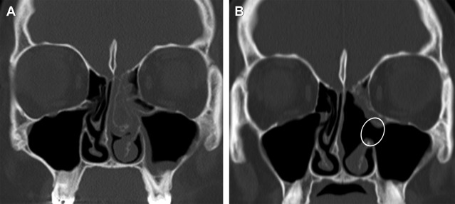
A possible case of symptomatic hemicrania continua from an osteoid osteoma  of the ethmoid sinus - KS Kim, HS Yang, 2010
A possible case of symptomatic hemicrania continua from an osteoid osteoma of the ethmoid sinus - KS Kim, HS Yang, 2010

Radio Scanner des sinus, OPN (os du nez) - Paris 18,17,9 - 92,93,94,95-  Doctolib
Radio Scanner des sinus, OPN (os du nez) - Paris 18,17,9 - 92,93,94,95- Doctolib

Radio Scanner des sinus, OPN (os du nez) - Paris 18,17,9 - 92,93,94,95-  Doctolib
Radio Scanner des sinus, OPN (os du nez) - Paris 18,17,9 - 92,93,94,95- Doctolib

Comment se passe un examen des sinus? - JeVaisMieuxMerci.com

The Preoperative Sinus CT: Avoiding a “CLOSE” Call with Surgical  Complications | Radiology
The Preoperative Sinus CT: Avoiding a “CLOSE” Call with Surgical Complications | Radiology

PDF] Radio anatomical analysis of positional relation between anterior  ethmoid artery canal and ethmoid skull base in correlation with olfactory  fossa | Semantic Scholar
PDF] Radio anatomical analysis of positional relation between anterior ethmoid artery canal and ethmoid skull base in correlation with olfactory fossa | Semantic Scholar
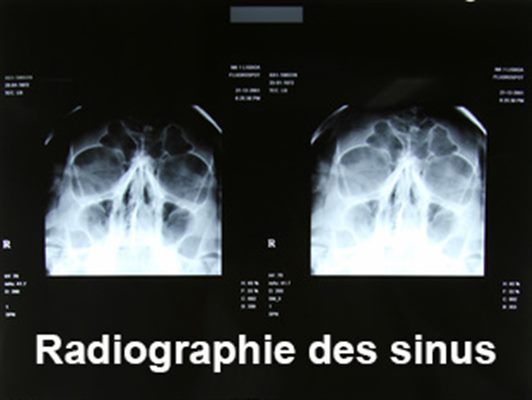
Radiographie des Sinus - Définition & Déroulement - Prise de rdv en ligne

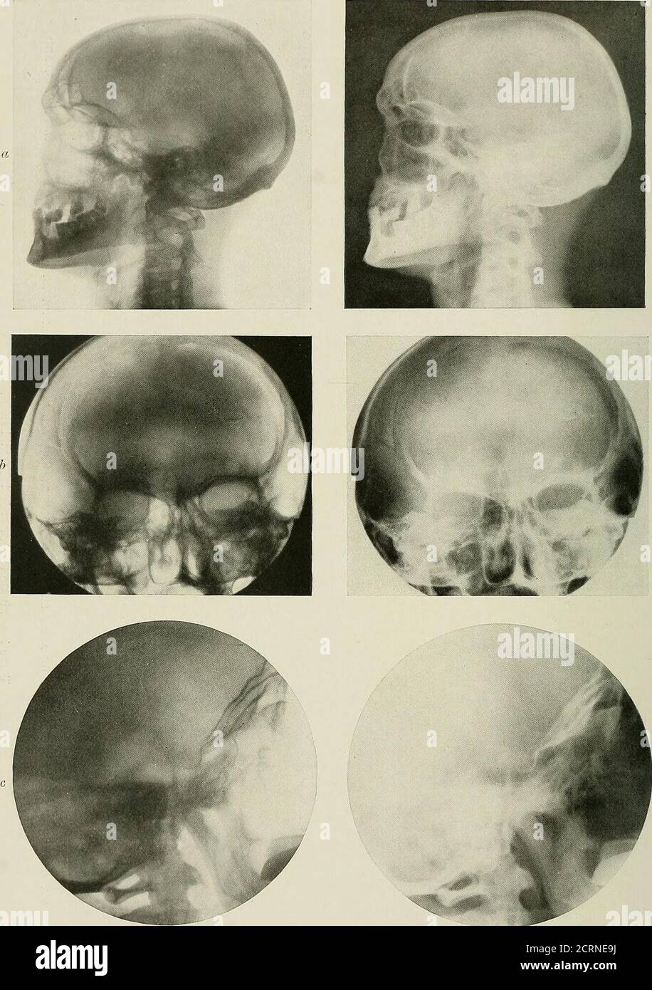
Radiography and radio-therapeutics . O ca a <. PLATE XV.—Normal Skdlls.  a, Lateral view of normal skull, showing frontal sinuses, sphenoidal  sinuses, sella turcica, temporal bones,cervical vertebras, and lower jaw. b,
Radiography and radio-therapeutics . O ca a <. PLATE XV.—Normal Skdlls. a, Lateral view of normal skull, showing frontal sinuses, sphenoidal sinuses, sella turcica, temporal bones,cervical vertebras, and lower jaw. b,

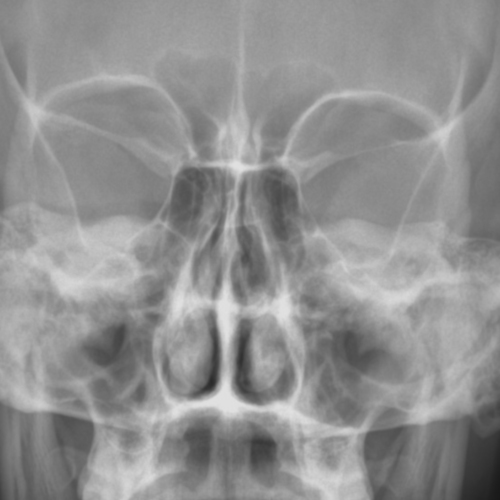
Radiographies des sinus examen - docteurclic.com

PNS radiograph revealing well defined radio-opaque shadow occupying the...  | Download Scientific Diagram
PNS radiograph revealing well defined radio-opaque shadow occupying the... | Download Scientific Diagram

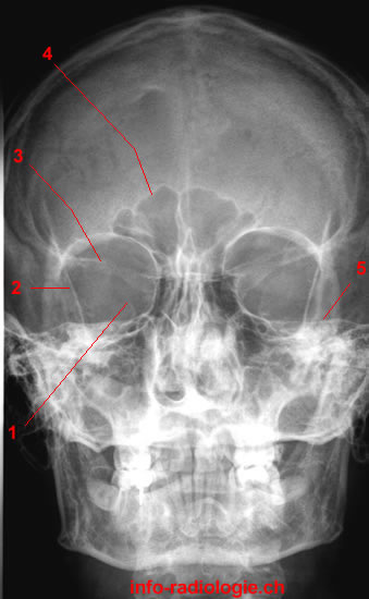
Radiographie des sinus de la face – Média LAROUSSE

CBCT showing right and left maxillary sinus radio-opacity CBCT:... |  Download Scientific Diagram
CBCT showing right and left maxillary sinus radio-opacity CBCT:... | Download Scientific Diagram

Oral medicine: 17. radiolucencies and radio-opacities. d. antral disease |  Dental Update
Oral medicine: 17. radiolucencies and radio-opacities. d. antral disease | Dental Update

Posttreatment Imaging of the Paranasal Sinuses Following Endoscopic Sinus  Surgery | Radiology Key
Posttreatment Imaging of the Paranasal Sinuses Following Endoscopic Sinus Surgery | Radiology Key

Figure 1 from Foreign Body in the Sphenoid Sinus | Semantic Scholar

Paranasal Sinuses Radiography - W-Radiology

The anterioposterior and lateral view X-ray showing the radio opaque... |  Download Scientific Diagram
The anterioposterior and lateral view X-ray showing the radio opaque... | Download Scientific Diagram
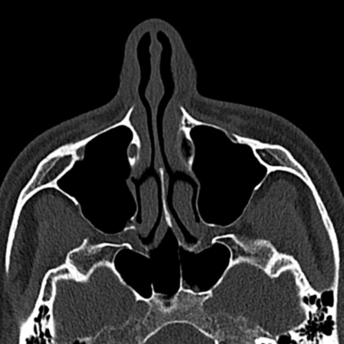
Ostiomeatal complex | Radiology Reference Article | Radiopaedia.org

The Preoperative Sinus CT: Avoiding a “CLOSE” Call with Surgical  Complications | Radiology
The Preoperative Sinus CT: Avoiding a “CLOSE” Call with Surgical Complications | Radiology

Stream Sinus #1 by Res.Radio | Listen online for free on SoundCloud

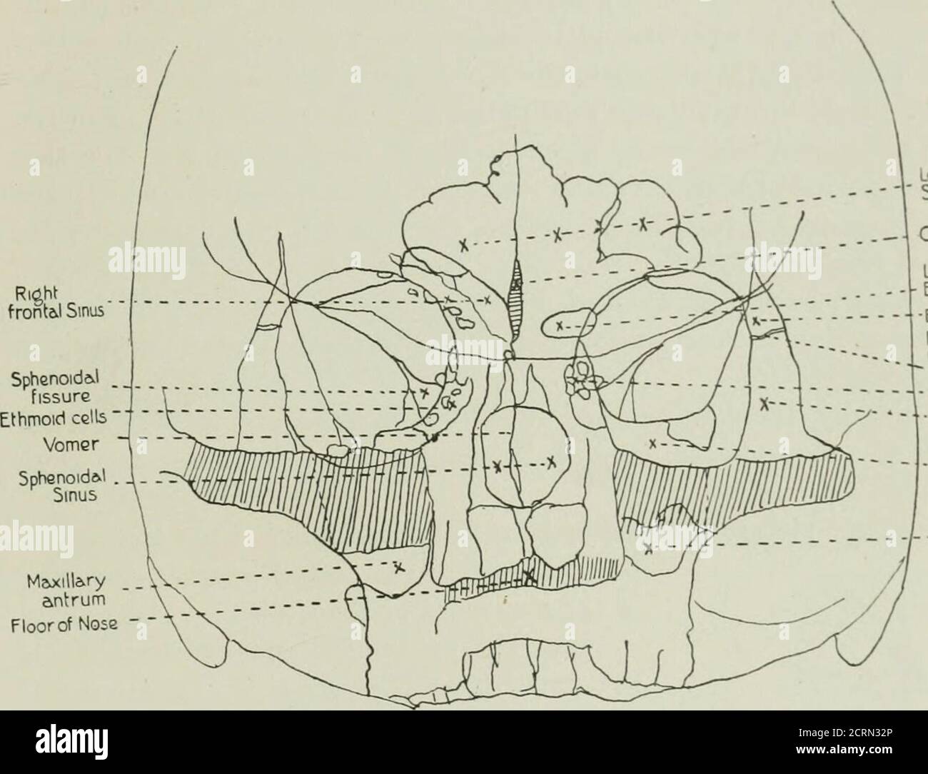
Radiography and radio-therapeutics . | inches above the external auditory  meatus, and passestowards the centre of the orbit. If the position is  correctly estimated the optic foramen will occupy thecentre of
Radiography and radio-therapeutics . | inches above the external auditory meatus, and passestowards the centre of the orbit. If the position is correctly estimated the optic foramen will occupy thecentre of

SiriusXM: Everything You Want to Hear Lives Here

Large central osteoma of maxillary sinus: A case report - ScienceDirect

Radiographies des sinus examen - docteurclic.com